
Pulmonary
In-vivo and functional assays were developed to study effects of drugs on pulmonary function
Technology
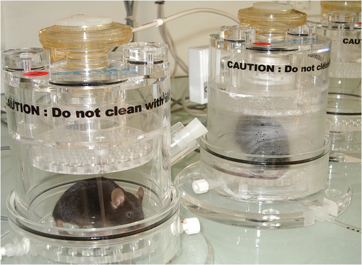
- Non invasive whole body plethysmography
- Sedated animal plethysmography and Flexivent®
Process 1: Whole body plethysmography
Process 2 : Airway resistance measurement on sedated animals
Whole body plethysmography analyzes can be confirmed by accurate measurements of resistance and compliance values of the airways in response to the administration of inhaled aerosolized cholinergic solutions by invasive plethysmography or Flexivent®.
- Airway resistance
- Dynamic Compliance
Functional tests :
- Flow cytometry analysis of Cell’s from broncho-alveolar lavage, lung, spleen and lymph nodes
- Complete blood count, analysis of immune serum globulins
- Morphological modification analysis on histological lung slides
- Pulmonary permeability (VLS)
- Effects of molecule on respiratory activity after nebulization
Animal Models:
Studies in animal models form the basis for much of our current understanding of the pathophysiology of asthma. Allergic mouse models of asthma are generated by sensitizing an animal to a foreign allergen.
Models:
Asthmatic mice are sensitized to an allergen, with or without adjuvant. After the immune system education which takes several days, the animals receive antigen exposure directly to the lungs via nasal instillation.
This elicits an inflammatory reaction in the lungs characterized essentially by an influx of eosinophils, and airways hyper-responsiveness (AHR) mimicking asthmatic symptoms.
We develop different protocols:
- Acute asthma models: study of asthma exacerbations (ovalbumine or house dust mite)
It takes about 4 weeks (depending on protocol) and allows the generation of effectors T cells in asthma (no clinical symptom appears during this phase). This leads to the generation of inflammation when animals receive antigen exposure directly to the lungs via nasal instillation.
- Chronic asthma models for the study of airway remodeling in asthma (ovalbumine or house dust mite)
These protocols take about 16 weeks (depending on protocol) and do not lead to any clinical symptoms. Mice are sensitized the first 3 weeks with allergen. Then during the following weeks, mice are regularly anesthetized to receive antigen exposure directly to the lungs via nasal instillation.


